Pesquisar neste blogue

quinta-feira, 30 de junho de 2011

Parafusos Absorvíveis para Reconstrução de Ligamento Cruzado Anterior (LCA)

Autores: Avelar SOS, Bretas CG, Carvalho LMA,
Kelles SMB, Mendonça ICA, Talim MCT, Alonso J , Cantanheda CRO, Conte T, Furlan LHP , Oliveira CAC, Pagnoncelli A , Menezes VM , Dr. Alexandre Pagnoncelli, Dr Carlos Augusto Cardim de Oliveira, Dra Claudia Regina de O. Cantanheda, Dra. Izabel Cristina Alves Mendonça, Dr. Jurimar Alonso.

Introdução - A reconstrução do ligamento cruzado anterior, por via artroscópica, é um procedimento ortopédico comum, em que se utiliza enxerto de tendão patelar ou de tendão semitendinoso ou grácil. A fixação destes enxertos é feita com o uso de parafusos de interferência metálicos ou bioabsorvíveis.
Os parafusos de interferência bioabsorvíveis foram introduzidos na década de 1990, tendo como vantagem descrita em relação aos parafusos metálicos, a possibilidade de facilitar uma eventual revisão cirúrgica, já que, uma vez absorvidos, dispensariam a etapa de remoção do parafuso. Seriam mais adequados, também, nos procedimentos de acompanhamento radiológico no pós-operatório, por não provocarem artefatos à ressonância magnética.
Além disto, poderiam representar menores riscos de laceração do enxerto com a mesma capacidade de fixação.

Objetivos – Estabelecer, através de revisão da literatura, se os benefícios esperados se confirmaram ao longo dos anos.

Métodos – Foi realizada busca no Medline, via PubMed, de revisões sistemáticas e ensaios clínicos randomizados publicados nos últimos cinco anos. Buscou-se, também, levantar séries de casos e relatos de casos, com o objetivo de se conhecer possíveis efeitos adversos relacionados a esta técnica.

Resultados – Foram encontrados uma revisão sistemática e cinco ensaios clínicos randomizados publicados nos últimos cinco anos, com amostras pequenas, mas com resultados consistentes. Um ensaio clínico não randomizado mostrou resultados semelhantes aos randomizados. Os relatos de casos servem como alerta para a possibilidade de efeitos adversos, que geralmente não são demonstrados nos ensaios clínicos.

Conclusão – Os estudos não demonstram benefícios clínicos ou funcionais adicionais dos parafusos absorvíveis. Houve ainda maior ocorrência de dor com a utilização destes, e as vantagens em relação à simplificação da técnica em casos de revisão são questionáveis, pelo lento processo de degradação e pelo processo inflamatório causado (cistos, pseudo-abscessos e migração dos parafusos). Não há evidências que justifiquem o uso preferencial
rotineiro dos sistemas de fixação absorvíveis ou que justifiquem seus custos
superiores.

Recomendação – Tendo em vista que os estudos clínicos randomizados comparando as técnicas de fixação de enxerto com parafusos absorvíveis e metálicos, não demonstraram superioridade dos parafusos absorvíveis em relação a desfechos clínicos e que a relação custo-efetividade ainda é desconhecida em nosso meio, a Câmara Técnica Nacional de Medicina Baseada em Evidências NÃO recomenda a incorporação dos sistemas de fixação absorvíveis em pacientes submetidos à reconstrução de ligamento cruzado anterior.


1. Questão Clínica

a) Há benefícios adicionais em termos de segurança e eficácia no uso de Parafusos Bioabsorvíveis para fixação femoral em Reconstrução de LCA com tendão patelar ou tendões flexores comparado ao uso de parafusos não absorvíveis?

b) Há benefícios adicionais em termos de segurança e eficácia no uso de Parafusos Bioabsorvíveis para fixação tibial em Reconstrução de LCA com tendão patelar ou tendões flexores comparado ao uso de parafusos nãoabsorvíveis?

2. Introdução

2.1. Aspectos epidemiológicos

A função do ligamento cruzado anterior (LCA) é a prevenção do deslocamento anterior da tíbia. O LCA costuma ser o ligamento mais comumente lesado e estar associado com o dano ao menisco ou com o ligamento medial colateral.


O sinal clínico característico da ruptura do LCA é o sinal da “gaveta” positivo, quando o joelho é flexionado a 90 graus e tracionado anteriormente, apresentando importante aumento da amplitude de movimento da articulação.


O LCA é o ligamento responsável pela estabilidade rotacional do joelho.
A lesão pode ocorrer após uma entorse do joelho, sendo mais comum durante atividade esportiva. Os sintomas principais da lesão são dor, derrame articular no joelho e impotência funcional. O tratamento na fase aguda consiste de antiinflamatórios, gelo, repouso e fisioterapia. O paciente com o LCA lesado consegue correr em linha reta, andar, nadar, porém ao praticar atividade na qual necessite "girar" sobre o joelho, vai sentir o joelho instável e terá grande chance de vir a ter novos entorses.
O manejo cirúrgico costuma ser indicado para pacientes com instabilidade articular e prejuízo de atividades diárias.
A reconstrução artroscópica do ligamento cruzado anterior (LCA) consiste no procedimento ortopédico cirúrgico mais comum atualmente.
Alguns tipos de enxertos têm sido utilizados na reconstrução do LCA como: o tendão patelar, os tendões flexores do joelho (semitendíneo e grácil) também chamados de isquiotibiais ou semitendinosos (“hamstring technique”), tendão quadriciptal ou tendão de cadáver 1.

As técnicas que utilizam o tendão patelar ou os tendões flexores proporcionam resultados clínicos e funcionais semelhantes, como foi demonstrado em vários ensaios clínicos randomizados. Estes resultados são satisfatórios em 85 a 90% dos pacientes 2,3,4,5,6,7.
Entretanto, no que se refere às técnicas de fixação do enxerto ainda existe alguma discussão.
A técnica mais comum de fixação utiliza parafusos de interferência metálicos, que podem ser usados tanto para fixação de enxertos patelares quanto nos semitendinosos 8.
As vantagens dos parafusos de interferência metálicos incluem fixação efetiva e confiável dos enxertos próximos à linha articular, com redução dos riscos de movimentação do enxerto dentro dos túneis ósseos e do alargamento destes túneis. 9,10
Desvantagens potenciais destes parafusos têm sido descritas como laceração das suturas, do enxerto e perda do plug ósseo durante a inserção. 11, 12, 13 Mas, a principal razão pela qual o uso dos parafusos metálicos nas intervenções de reconstrução do LCA tem sido questionado foi a dificuldade de remoção dos mesmos nos eventos de revisão cirúrgica, e os possíveis artefatos provocados nas imagens de ressonância magnética, quando há necessidade deste tipo de avaliação no pós-operatório. 14
Estes fatores levaram ao interesse crescente pelos parafusos de interferência bioabsorvíveis, teoricamente capazes de superar as dificuldades acima descritas relacionadas aos parafusos metálicos, com a mesma capacidade de fixação do enxerto. Estes parafusos foram introduzidos nos anos 1990 e, segundo os fabricantes, seriam completamente degradados e absorvidos até dois anos após o implante, permitindo a reposição do tecido ósseo nos sítios tibial e femoral de implante dos mesmos.
Entretanto, a velocidade de absorção destes parafusos depende de uma série de fatores, que incluem a composição dos mesmos, massa, quantidade de cristalização, cobertura da superfície e o ambiente em que são inseridos. 15

2.2. Descrição da tecnologia

Os parafusos de interferência bioabsorvíveis são constituídos por polímeros de ácido poliglicólico (PGA) ou de ácido poliláctico (PLA). Alguns representam associação destes dois ácidos, enquanto outros têm um destes ácidos associados a outros polímeros ou a materiais biocerâmicos como a Hidroxiapatita (HA) ou o fosfato tricálcico (TCP). Geralmente, o ácido poliglicólico tem um processo de absorção mais rápido que o ácido polilático. A absorção destes polímeros pelo organismo é feita através de hidrólise lenta, com liberação, ao final, de CO2 e água.
O processo de degradação no organismo de qualquer um destes materiais não é dependente de atividade enzimática 16, mas pode provocar reações inflamatórias inespecíficas com ativação de fibroblastos, macrófagos e leucócitos polimorfonucleares. Este processo pode durar até cinco anos. 16,17
Existem diversos modelos de sistemas de fixação de tendão absorvíveis sendo comercializados em nosso país para fixação tibial e femoral.

TIBIAIS

 FEMURAIS
TIBIAIS E FEMURAIS

Como alternativa existem os sistemas de fixação de enxertos nãoabsorvíveis como os parafusos de interferência metálicos. Alguns modelos em titânio vêm sem bordas, o que previne a laceração do enxerto e não interfere com a resolução das imagens radiológicas à ressonância magnética.
Além destes são comercializados também outros sistemas de fixação não-absorvíveis como parafusos com arruelas em titânio ou aço, pinos transversais em titânio, endobottons em titânio, grampos em titânio e parafusos em polietileno.

TIBIAIS


3. Metodologia

3.1. Bases de dados pesquisadas:

Identificação de avaliações da tecnologia já realizadas por Câmaras Técnicas do Sistema Unimed, Medline via Pubmed, Cochrane, Agências de Avaliação de Tecnologia da INAHTA .

3.2. Palavras-chave ou Descritores (DeCS) utilizados: anterior cruciate ligament, bone screws, absorbable implants, intrafix, rigidifix.

3.3. Desenhos dos estudos procurados:
Metanálises e Ensaios Clínicos Randomizados ou não randomizados.
Na ausência destes, buscou-se também, levantar séries de casos e relatos de casos, com o objetivo de se conhecer possíveis efeitos adversos relacionados a esta técnica.

3.4. População envolvida:
Pacientes submetidos à reconstrução de ligamento cruzado anterior (LCA)

3.5. Intervenção:
Fixação dos enxertos com sistemas de fixação absorvíveis.

3.6. Comparação:
Fixação dos enxertos com sistemas de fixação metálicos.

3.7. Desfechos:

Clínicos (funcionalidade, infecção, dor); desfechos substitutos (laceração de suturas e do enxerto, dificuldade de remoção do implante em revisão cirúrgica, artefatos provocados nas imagens de ressonância magnética no pós-operatório, taxas de reintervenção), alargamento de túneis ósseos, tempo de absorção do parafuso.

3.8. Período da pesquisa:

Últimos cinco anos.
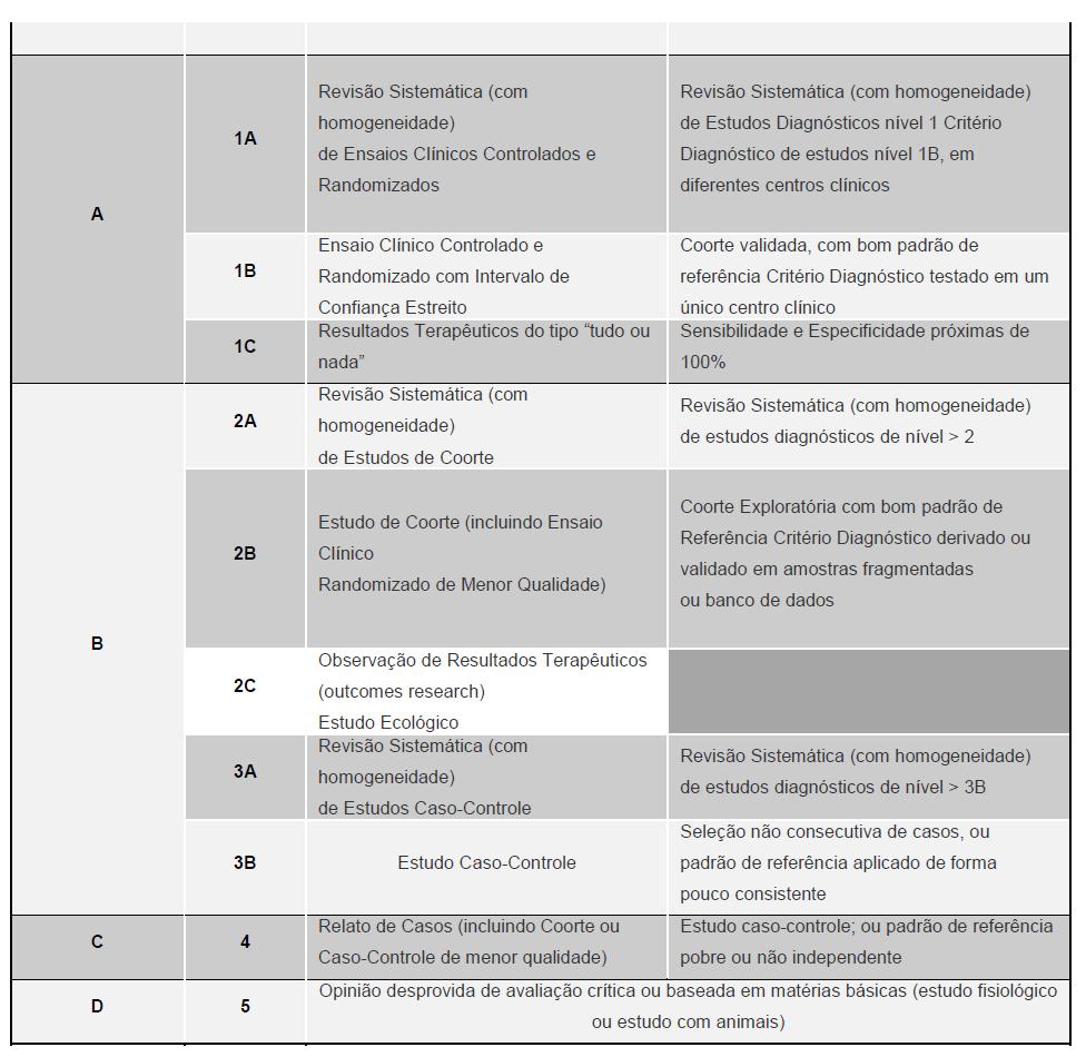
O grau de recomendação tem como objetivos dar transparência às informações, estimular a busca de evidência científica de maior força e auxiliar a avaliação crítica do leitor, o responsável na tomada de decisão junto ao paciente.


4. Resultados

4.1. Comentários

Os resultados, quanto a desfechos clínicos, após a reconstrução cirúrgica do ligamento cruzado anterior são semelhantes, tanto na fixação tibial quanto na femoral, com os dois tipos de fixação utilizados, ou seja, com parafusos de metal ou com parafusos bioabsorvíveis, como já havia sido demonstrado na revisão feita pelos grupos de avaliação de tecnologias do Sistema Unimed 24, 25 em 2005 e 2006 e confirmado pelos ensaios clínicos randomizados mais recentes 20,21,22. Estes ensaios, embora individualmente com amostras pequenas, demonstram resultados bastante semelhantes e, portanto, são consistentes. São ensaios com critérios de exclusão rígidos, incluindo geralmente pacientes com acometimento restrito ao ligamento cruzado anterior, unilateralmente.
As possíveis vantagens do parafuso bioabsorvível quanto à facilitação de revisões cirúrgicas, pela não necessidade de remoção do parafuso são questionáveis, uma vez que a degradação e absorção do parafuso são lentas, durando cerca de quatro anos. Assim, qualquer revisão durante este longo período, necessitará da retirada do parafuso.
Por outro lado, a absorção, mesmo completa do parafuso, não foi acompanhada de reconstituição óssea nos locais onde os mesmos foram colocados 16.
A possibilidade de avaliação através de ressonância magnética no pós-operatório, outro fator apontado como vantagem relacionada ao uso de parafusos de interferência bioabsorvíveis, também pode ser questionada porque resultados semelhantes podem ser obtidos com os parafusos de titânio, que também não prejudicam a avaliação por este método radiológico.
A ressonância magnética, ainda que precisa para verificar o estado do enxerto, pode não ter a mesma precisão em determinar a absorção do parafuso e sua eventual substituição por tecido ósseo, como demonstrado no relato de caso feito por Park e Tibone 17. Estes autores descrevem um caso de nova abordagem cirúrgica motivada por dor e edema sobre a região anterior da tíbia, mais de quatro anos após a reconstrução do LCA. A avaliação através da ressonância no pré-operatório indicava que o parafuso bioabsorvível usado na primeira cirurgia estava intacto. No per operatório constatou-se que não havia resquício do parafuso, sendo curetado material amorfo, compatível com abscesso estéril.
O uso de parafusos biodegradáveis na fixação do enxerto tendíneo seja ele patelar ou semitendinoso, não está isenta de riscos. Como o caso acima mencionado, há descrição de formação de abscessos estéreis pré-tibiais, a partir de cistos, tardiamente após a reconstrução do ligamento cruzado anterior 26. A migração do parafuso, uma complicação inicialmente creditada apenas aos parafusos de interferência metálicos, também foi descrita com parafusos bioabsorvíveis 27,28, sobretudo com aqueles usados para a fixação tibial. A migração do parafuso geralmente é uma intercorrência precoce que ocorre dentro dos primeiros meses após a cirurgia e é causa de dor e de limitação dos movimentos do joelho.
As evidências de que não há benefício clínico ou funcional adicional dos parafusos de interferência bioabsorvíveis em relação aos parafusos metálicos são consistentes e robustas.
As vantagens quanto à simplificação técnica nos casos de revisão cirúrgica são questionáveis diante do curso muito lento do processo de degradação e absorção dos parafusos bioabsorvíveis, implicando que, em muitas revisões cirúrgicas, ocorra a mesma necessidade de remoção destes parafusos que ocorre com os parafusos metálicos. Além disto, o processo lento de absorção implica em reação inflamatória com riscos de formação de cistos, pseudo-abscessos e migração dos parafusos.
Os possíveis benefícios advindos da ausência de artefatos aos exames de ressonância magnética também podem ser usufruídos com o uso de parafusos de titânio.
Não foram encontrados estudos clínicos em humanos comparando o sistema de pinos transversos absorvíveis (Rigidifix ®) ou o sistema de fixação com parafuso e bainha, em polietileno ou absorvível (Intrafix ®) com sistemas de fixação metálicos.

4.2. Avaliação de custos

Os sistemas de fixação ligamentar absorvíveis ou não, apresentam grande variação de preços no mercado.


Relação de Registros na Anvisa e Cotação de Preços

5. Conclusão e recomendações

5.1. Conclusão

Os estudos demonstram os seguintes resultados do emprego de sistemas de fixação absorvíveis para reconstrução de ligamento cruzado anterior em relação ao emprego de sistemas de fixação metálicos:
Não há benefícios clínicos adicionais 18, 20, 21, 29 ;
Não há benefícios funcionais adicionais 18, 19, 22, 23, 29 ;
As evidência são controversas em relação ao retorno à atividade física 19, 20;
Há registros de maior ocorrência de dor com o uso de parafusos absorvíveis 19, 20;
As vantagens em relação à simplificação da técnica em casos de revisão são questionáveis pelo lento processo de degradação e pelo processo inflamatório causado (cistos, pseudo-abscessos e migração dos parafusos) 15, 16, 17, 23;
Não há evidências que justifiquem o uso preferencial rotineiro dos sistemas de fixação absorvíveis ou que justifiquem seus custos superiores;

5.2. Recomendação da Câmara Técnica Nacional de Medicina Baseada em Evidências (CTNMBE):

Tendo em vista que os estudos clínicos randomizados comparando as técnicas de fixação de enxerto com parafusos absorvíveis e metálicos, não demonstraram superioridade dos parafusos absorvíveis em relação a desfechos clínicos e que a relação custo-efetividade ainda é desconhecida em nosso meio, a Câmara Técnica Nacional de Medicina Baseada em Evidências NÃO recomenda a incorporação dos sistemas de fixação absorvíveis em pacientes submetidos à reconstrução de ligamento cruzado anterior.


6. Referências Bibliográficas:

1. Sociedade Brasileira de Cirurgia de Joelho. Enquete. Disponível em
http://www.sbcj.org.br/sbcj.cfm?cmt=1&p1_enq=1461429&categ_id=1208885&env=1 .
Acesso em 06/12/2008.

2. Aglietti P, Giron F, Buzzi R, Biddau F, Sasso F. Anterior cruciate ligament reconstruction: bone-patellar tendon-bone compared with double semitendinosus and gracilis tendon grafts. A prospective, randomized clinical trial. J Bone Joint Surg Am. 2004 Oct;86-A(10):2143-55.

3. Beynnon BD, Johnson RJ, Fleming BC, Kannus P, Kaplan M, Samani J, Renström P.
Anterior cruciate ligament replacement: comparison of bone-patellar tendon-bone grafts with two-strand hamstring grafts. A prospective, randomized study. J Bone Joint Surg Am. 2002 Sep;84-A(9):1503-13.

4. Ejerhed L, Kartus J, Sernert N, Köhler K, Karlsson J. Patellar tendon or semitendinosus tendon autografts for anterior cruciate ligament reconstruction? A prospective randomized study with a two-year follow-up. Am J Sports Med. 2003 Jan-Feb;31(1):19-25.

5. Eriksson K, Anderberg P, Hamberg P, Löfgren AC, Bredenberg M, Westman I, Wredmark T. A comparison of quadruple semitendinosus and patellar tendon grafts in reconstruction of the anterior cruciate ligament. : J Bone Joint Surg Br. 2001 Apr;83(3):348-54.

6. Jansson KA, Linko E, Sandelin J, Harilainen A. A prospective randomized study of patellar versus hamstring tendon autografts for anterior cruciate ligament reconstruction. Am J Sports Med. 2003 Jan-Feb;31(1):12-8.

7. Laxdal G, Kartus J, Hansson L, Heidvall M, Ejerhed L, Karlsson J. : A prospective randomized comparison of bone-patellar tendon-bone and hamstring grafts for anterior cruciate ligament reconstruction. Arthroscopy. 2005 Jan;21(1):34-42.

8. Kurosaka M, Yoshiya S, Andrish JT. A biomechanical comparison of different surgical techniques of graft fixation in anterior cruciate ligament reconstruction. Am J Sports Med. 1987;15:225-229.

9. Paschal SO, Seemann MD, Ashman RB, Allard RN, Montgomery JB. Interference fixation versus postfixation of bone–patellar tendon– bone grafts for anterior cruciate ligament reconstruction: a biomechanical comparative study in porcine knees. Clin Orthop Relat Res. 1994;300:281-287.

10. Clatworthy MG, Annear P, Bulow JU, Bartlett RJ. Tunnel widening in anterior cruciate ligament reconstruction: a prospective evaluation of hamstring and patella tendon grafts. Knee Surg Sports Traumatol Arthrosc. 1999;7:138-145.

11. Bach BB. Potential pitfalls of Kurosaka screw interference fixation for ACL surgery. Am J Knee Surg. 1989;2:76-82.

12. Lemos MJ, Jackson DW, Lee TQ, Simon TM. Assessment of initial fixation of endoscopic interference femoral screws with divergent and parallel placement. Arthroscopy. 1995;11:37-41.

13. Pierz K, Baltz M, Fulkerson J. The effect of Kurosaka screw divergence on the holding strength of bone-tendon-bone grafts. Am J Sports Med. 1995;23:332-335.

14. Shellock FG, Mink JH, Curtin S, Friedman MJ. MR imaging and metallic implants for anterior cruciate ligament reconstruction: assessment of ferromagnetism and artifact. J Magn Reson Imaging. 1992;2:225-228.

15. Radfort MJ, Noakes J, Read J, Wood DG. The natural history of a bioabsorbable interference screw used for anterior cruciate ligament reconstruction with a 4-strand hamstring technique. Arthroscopy 2005;21(6): 707-710.

16. Barber FA, Dockery WD. Long-term absorption of poly-L-lactic Acid interference screws. Arthroscopy 2006;22(8): 820-6.

17. Park MC, Tibone JE. False magnetic resonance imaging persistence of a
biodegradable anterior cruciate ligament interference screw with chronic inflammation
after 4 years in vivo. Arthroscopy 2006;22(8): 911.e1-4.

terça-feira, 28 de junho de 2011

Improving Walking After Stroke using a trendmill

Autores: LOUISE ADA AND CATHERINE M. DEAN


CASE REPORT I

BACKGROUND

Mrs PG is 65 and lives with her husband, who is still working part time. She woke up not being able to speak coherently and not being able to move the right side of her body. The ambulance was called and she was admitted to hospital. It is now Day Six.

MEDICAL STATUS

Diagnosed having had a stroke. Conscious. On blood pressure lowering medication.

IMPAIRMENTS

Weakness – severe in most lower limb muscles and all upper limb muscles.

Incoordination – unable to be assessed due to severe weakness.

Spasticity – no spasticity – Tardieu scale score X = 0 at V3 (fast velocity) during ankle dorsiflexion and elbow extension.

Sensation – normal.

Language – expressive aphasia so she understands 90 % but can only communicate about 40 % of what she wants to say.

Cognition – normal.
Perception – normal.

ACTIVITY LIMITATIONS

Standing – cannot stand independently, needs help from one person.
Walking – cannot walk independently, needs substantial help from two people.

Use of upper limb – no voluntary movement at any joint.

QUESTION 1


Should treadmill training with body weight support (BWS) be used to retrain walking?
The first step in answering this question is to decide on the outcomes of interest.
Given that Mrs PG is non-ambulatory, whether or not treadmill training with BWS is effective at establishing walking will be of prime interest. Furthermore, the quality of the walking produced by the training will be of interest. There are numerous outcome measures which evaluate walking, ranging from performance-based tests (such as the 10-m Walk Test (Wade 1992)) to ordinal scales (such as Item 5 of the Motor Assessment Scale for Stroke (Carr et al. 1985)). The most commonly used measure in clinical trials is the 10-mWalk Test, probably because it is simple to carry out, reliable, and yields continuous data. Furthermore, the most common parameter reported is walking speed and, while not measuring quality of walking directly, it nevertheless reflects qualitative gait parameters such as step length and cadence. This relationship is described in the equation:

Speed = (stride length × cadence)/120

Therefore, proportion of patients walking and the 10-m Walk Test are probably the best measures reflecting the outcomes of interest.
The next step in answering the question is to look for evidence of whether treadmill training improves the proportion of people walking independently, or the quality of walking. Considering the highest levels of evidence first, there are two systematic reviews assessing the efficacy of treadmill training with BWS after stroke. The efficacy of treadmill training with BWS was considered in a review by Van Peppen and colleagues (2004 A). They concluded that treadmill training with BWS does not improve walking speed or ability although it does appear to improve walking endurance.
However, most of the participants in the trials included in this review were already walking and so this finding is of limited use in answering the question. The efficacy of treadmill training with BWS was also considered in a Cochrane review by Moseley and colleagues (2005 A). They did separate their analyses into those who were non-ambulatory versus those who were ambulatory. They report that there is no greater risk of being non-ambulatory or a dependent walker if treadmill training with BWS is used than if other more conventional interventions are used (RR 1.1, 95 % CI 0.9 to 1.3). This finding was based on 178 participants in five randomised trials (da Cunha Filho et al. 2002, Kosak et al. 2000, Nilsson et al. 2001, Scheidtmann et al. 1999, Werner et al. 2002 A). Furthermore, walking speed was no different as a result of the interventions (WMD –0.01 m/s, 95 % CI –0.08 to 0.06). This finding was based on 148 participants in four randomised trials (da Cunha Filho et al. 2002; Kosak et al. 2000; Nilsson et al. 2001;Werner et al. 2002 A). The more conventional therapy used in these trials was always exactly matched for frequency and duration and was usually carried out for 20–45 minutes, five days a week. Two trials used a motor learning approach (da Cunha Filho et al. 2002; Nilsson et al. 2001 A), while one trial used a neurophysiologic approach (Scheidtmann et al. 1999), one used an orthopaedic approach (Kosak et al. 2000 A), and one used another walking device  (Werner et al. 2002 A).
Given that treadmill walking with BWS is no more or less effective than the same amount of conventional therapy, the decision of whether to undertake it with Mrs PG will have to be made on other factors. Such factors are the efficient use of staff time, the amount of practice likely to be undertaken during overground walking versus treadmill walking, and Occupational Health and Safety. At the moment, it takes two therapists to help Mrs PG practise the whole task of walking overground. While this was controlled in the randomised trials, it is unlikely two staff members will be free to help her for very long in ordinary clinical practice. Walking on the treadmill with BWS means that she may only need the help of one therapist to move her affected leg forward in swing phase, even if it takes two people to assist her onto the treadmill. It is likely to be easier to move Mrs PG’s leg during swing when she is in one place on a treadmill than to support it during swing and stance while she is trying to progress overground (since it does not matter if the knee flexes during stance, as the body is supported). Treadmill walking with partial weight support via an overhead harness provides the opportunity to complete larger amounts ofwalking practice, for example, even if patients only walk for five minutes at a slow speed of 0.2 m/s supported on a treadmill, they will ‘walk’ 60 m (Crompton et al. 1999 C). It is likely, therefore, that Mrs PG will undertake more practice of the whole task of walking if she does treadmill training with BWS.
Taking into account all the evidence, treadmill training with BWS should be an intervention capable of establishing walking in Mrs PG.

QUESTION 2

How should treadmill training with BWS be applied to improve the likelihood of the patient becoming ambulatory with good quality of walking?

To answer this question, observational studies of treadmill and overground walking after stroke can be examined. These studies compare walking overground with walking on a treadmill with BWS in stroke patients who are just walking or walking with difficulty (Chen et al. 2005a, 2005b; Hassid et al. 1997; Hesse et al. 1997 A). One of the common findings is that by adding BWS, the symmetry of walking is improved, due to the increased time the affected leg spends in single stance phase. However, there may be a limit to how much support should be given. Hesse and colleagues (1997 A) compared 0, 15, 30, 45 and 60 % BWS. They found that over 30 % BWS resulted in markedly abnormal muscle activity in six lower limb muscles they examined. This has resulted in a maximum of 30 % BWS becoming something akin to an industry standard. Perhaps the most useful information comes from Chen and colleagues (2005a, 2005b A), who systematically varied BWS, speed of treadmill, stiffness of the support harness, and support from a handrail. They found that different factors were helpful in different aspects of walking. For example, increasing BWS combined with support from a handrail produced the most symmetrical walking in orthopaedic approach (Kosak et al. 2000 A), and one used another walking device  (Werner et al. 2002 A).
Given that treadmill walking with BWS is no more or less effective than the same amount of conventional therapy, the decision of whether to undertake it with Mrs PG will have to be made on other factors. Such factors are the efficient use of staff time, the amount of practice likely to be undertaken during overground walking versus treadmill walking, and Occupational Health and Safety. At the moment, it takes two therapists to help Mrs PG practise the whole task of walking overground. While this was controlled in the randomised trials, it is unlikely two staff members will be free to help her for very long in ordinary clinical practice. Walking on the treadmill with BWS means that she may only need the help of one therapist to move her affected leg forward in swing phase, even if it takes two people to assist her onto the treadmill. It is likely to be easier to move Mrs PG’s leg during swing when she is in one place on a treadmill than to support it during swing and stance while she is trying to progress overground (since it does not matter if the knee flexes during stance, as the body is supported). Treadmill walking with partial weight support via an overhead harness provides the opportunity to complete larger amounts ofwalking practice, for example, even if patients only walk for five minutes at a slow speed of 0.2 m/s supported on a treadmill, they will ‘walk’ 60 m (Crompton et al. 1999 C). It is likely, therefore, that Mrs PG will undertake more practice of the whole task of walking if she does treadmill training with BWS.
If the affected arm has no voluntary muscle activity, use a firm sling to support it, but if there is some activity, put the hand to the handrail using a bandage or a weightlifting splint (see Figure 1).


We have found metronomes to be usefulin enhancing rhythmical stepping and thereby directing step length; for example, slowing the metronome down will result in alternate feet staying on the ‘ground’ for longer. The most difficult job for the therapist is to lift the affected leg through during swing phase (Figure 2a). When the leg is very weak, a length of theraband can be tied from the front of the shoe to the front bar of the treadmill, which will serve to pull the leg forward when the weight is released (Figure 2c). Alternately, the affected foot can be placed in a pillow slip and twisted at the front (Figure 2b) so that the foot can be lifted from the toe, thereby enhancing dorsiflexion of the ankle.


The therapist can sit on a chair turned backwards, which will support the trunk, making lifting the affected leg easier. It is important that the therapist assists the leg only in swing phase, and encourages the patient to extend their lower limb during stance, allowing the BWS to prevent the patient collapsing. To progress the training, when step length is consistently normal, we increase the speed until step length is compromised.
When the knee can be held straight during stance phase, we reduce the BWS.We have found  that an easy transition is made to overground walking when the patient can walk on the treadmill at 0.5 m/s with ≤ 10 % BWS.

PLAN: TO ESTABLISH GOOD QUALITY WALKING IN MRS PG

A specific intervention plan, based on the above evidence, to carry out treadmill training with BWS for Mrs PG using a treadmill and overgound BWS system, is outlined below:

Gain medical clearance and consent to participate in exercise programme Consult with Mrs PG’s treating doctor to organise medical clearance to participate in treadmill walking training with BWS. Put harness on in lying and make sure Mrs PG is wearing shoes. Apply triangular sling to affected arm. Wheel Mrs PG onto treadmill in a wheelchair. Use the automatic lift function to lift her into standing. Given that Mrs PG has communication problems, modified safety procedures will have to be put in place. Attach safety strap, have relative or aide standing by emergency stop switch and teach Mrs PG a signal to indicate that the treadmill should be stopped.

Initial treadmill and BWS programme

To begin with, do not run the treadmill. Allow Mrs PG to hang on to a handrail. Increase BWS to 30%in standing and make sure knee of affected leg is bent no more than 15 degrees. If it is, increase BWS. Put Mrs PG’s affected foot in a pillow slip and twist at the front. Turn on a metronome at a frequency which matches the highest  cadence that Mrs PG can manage. Sit on a low stool and help Mrs PG to walk on the spot in time with the rhythm by using the right hand to flex the knee and the left hand to lift the twisted part of the pillowcase. Then turn the treadmill on as slowly as possible. Mrs PG should keep walking in time with the metronome – the metronome frequency and the treadmill speed will determine her step length. Lift the affected leg forward during swing phase but encourage Mrs PG to extend her lower limb during stance and allow the BWS to hold her up. Count steps for encouragement and take a rest every two minutes at first.
Progressing treadmill and BWS programme Increase step length by slowing down the metronome. When step length is increased, increase the speed until step length is compromised. When Mrs PG can straighten her knee from the 15 degrees, reduce the BWS. Continue to alternate these two strategies until she is walking at 0.5 m/s with≤10%BWS. At this stage begin to do overground walking with BWS.

Overground and BWS programme

Lock the wheels of the support frame so that it will only run in one direction. Put markers on the floor to increase step length and constrain step width. Apply only the trunk/pelvis part of harness, firmly. Push the support frame as Mrs PG walks forwards and then backwards overground. Progress by loosening the vertical support straps, getting Mrs PG to push the frame herself, and increasing step length and decreasing step width (see Figure 3).

Monitoring progression and enhancing compliance
At the beginning, record the number of steps to provide encouragement. Then, as ability improves, record distance covered on treadmill, highest speed and lowest amount of BWS – graph to provide motivation to improve. Record distance, step length and width during overground walking with BWS. As independent walking overground is possible, use 10-m Walk Test at the same time every week to monitor progress. As well as timing over the 10-m, count the number of steps and calculate average step length and cadence.

CASE REPORT II

BACKGROUND

Mr IB is 70 and lives alone. He has a very supportive daughter nearby, although she is busy bringing up four children. He suffered a stroke two years ago. Recently he has felt that his walking has deteriorated, and has approached a physiotherapy ambulatory care service for help.

 
Figure 3. Using a portable system to practise overground walking. Harness is for safety  only. Markers on the floor encourage a long step length and narrow step width (refers to Case Report II).

MEDICAL STATUS

On blood pressure lowering medication.

IMPAIRMENTS

Weakness – moderately strong in lower limb muscles.

Incoordination – slight problem with incoordination in both upper limb and lower limb.

Spasticity – mild spasticity – Tardieu scale score X = 1 at V3 (fast velocity) during ankle dorsiflexion and X = 2 during elbow extension. Contracture – loss of 10◦ ankle dorsiflexion.

Sensation – tactile and kinaesthetic sensation moderately impaired.

Language – normal.

Cognition – slight memory loss.

Perception – normal.

ACTIVITY LIMITATIONS

Standing – can stand with feet together and look over both shoulders without falling or having to take a step, but uses arms.

Walking – can walk independently, but very slowly and carefully at 0.6 m/s and 190m in six min.

Use of upper limb – can use for support but not manipulation of objects (mostly due to loss of sensation).

In addition to the trials that match Mr IB’s characteristics, there is more evidence (although at a weaker level) which suggests treadmill walking may be a useful intervention to improve both the speed and capacity of walking in such patients. In uncontrolled trials of chronic stroke patients, treadmill walking has been associated with increases in strength (Smith et al. 1998, 1999 A), decreases in energy expenditure(Macko et al. 1997, 2001 A), as well as increases in walking speed and quality (Silver et al. 2000 A).
 Taking into account all the evidence, treadmill training should be an intervention capable of improving Mr IB’s community ambulation.

QUESTION 2


How should treadmill training be applied to improve community ambulation?
The most logical approach to answering this question is to examine how the intervention was implemented in the two trials which provided evidence that treadmill walking was effective in improving six minute distance (Ada et al. 2003; Macko et al. 2005 A). Macko and colleagues’ training programme was six months, 10–40 minute sessions, three times a week. The sessions were characterised by progressive increases in duration (five minutes per session every two weeks) and in aerobic intensity (5 % Heart Rate Reserve every two weeks, achieved by increasing the speed
of the treadmill). Training speed increased from 0.48 ± 0.3 m/s at baseline to 0.75 ± 0.3 m/s at six months, and training duration increased from 12 ± 6 minutes to 41 ± 10 minutes at six months.

Ada and colleagues implemented a training programme three times a week for only four weeks. The training sessions comprised 30 minutes of walking, which took about 45 minutes to accomplish. Each session consisted of both treadmill and overground walking, with the proportion of treadmill walking decreasing by 10 % each week, from 80%inWeek One to 50%inWeek Four. Subjects received individual training from a physical therapist; however, there was some opportunity for social interaction since two subjects were trained concurrently. The programme was carried out in a community setting and transport was provided if necessary. The treadmill walking componentwas structured to increase step length, speed, balance, fitness, and automaticity. To increase step length, the treadmill was run at a comfortable speed and instructions such as ‘walk as slowly as possible’ or ‘take as few steps as possible’ were used. When a normal step length was observed, the speed of the treadmill was increased (until step length was compromised). When maximum speed was achieved, balance was challenged by reducing the degree of hand support, and fitness encouraged by increasing the incline of the treadmill, thereby increasing workload.
Finally, automaticity was promoted by presenting the subjects with a concurrent cognitive task (Canning et al. 2006 A; Paul et al. 2005). The cognitive task consisted of matching the word ‘red’ with the response ‘yes’, or the word ‘blue’ with the response ‘no’ (Bowen et al. 2001 A).
The overground walking component aimed to reinforce improvements in walking pattern and speed achieved on the treadmill. To reinforce the increased step length, visual cues were used in the form of non-slip footprints, which were laid at intervals normal for that subject’s height. As step length approximated normal, subjects were encouraged to walk faster and were timed for feedback. Step width was reduced and balance challenged by forcing subjects to walk within one floor tile or walk along a line forwards, sideways and backwards. Workload was increased by introducing stairs and slopes to overground walking practice, and automaticity was promoted by the introduction of dual tasks. Subjects walked continuously around an outdoor circuit, which included curbs, slopes, stairs and rough terrain, while conversing with the trainer. The immediate improvement in walking capacity measured by the 6-min. Walk Test was greater in the Ada and colleagues study than in Macko and colleagues.
As described above there were differences in the programmes, which may account for these results. Macko and colleagues only used treadmill training, with increasing speed and session duration, whereas Ada and colleagues’ programme involved treadmill and overground walking, focusing not only on fitness but also on quality and automaticity of walking. There is other evidence to suggest that the content of a treadmill walking programme is important in determining effectiveness. For example, Pohl and colleagues (2002 A) have shown the importance of manipulating the speed of the treadmill to achieve increases in overground walking speed. However, it has been shown that stroke patients generally achieve higher walking velocities by increasing their cadence rather than step length (Wagenaar et al. 1992 A). We therefore suggest that treadmill training programmes should include overground walking components where increases in walking speed and step length are encouraged. The improvements in walking capacity were not maintained in the Ada and colleagues study, which suggests that the one month duration was insufficient and that treadmill programmes should be of longer duration, such as the six months used by Macko and colleagues.
Based on the strategies implemented by Macko and colleagues and Ada and colleagues, we would recommend a treadmill and overground programme of 30 to 40 minutes three times a week for four to six months, with training aimed to increase speed, step length, aerobic intensity and automaticity.

PLAN: TO IMPROVE MR IB’S COMMUNITY AMBULATION

Aspecific intervention plan for Mr IB, based on the above evidence, is outlined below:
Gain medical clearance and consent to participate in exercise programme Consult with Mr IB’s treating doctor to organise medical clearance or stress test (as per ASCM guidelines) to participate in a treadmill and overground walking programme aimed at improving walking capacity and aerobic fitness.
Clinical facility: supervised treadmill overground walking programme focused on improving step length

Arrange for Mr IB to attend ambulatory care/out-patient setting three times a week for two weeks. Negotiate with his daughter to provide transport or organise community transport. At the initial session, determine if other impairments are interfering with walking and if so recommend treatment or adaptation. Mr IB’s impaired sensation may be a reason he cannot walk backwards, since in this situation he has no peripheral vision of his feet. Commence a supervised treadmill and overground walking programme focusing on increasing step length and then increasing speed and step length. Include a warm up, involving stretches of the calf and hip flexor muscles against a  wall, before Mr IB gets on treadmill.
Local gym: treadmill training focusing on aerobic training

Consult with Mr IB and his daughter to find a local gym with a treadmill which Mr IB can access without relying on her for transport. Use a heart rate monitor (pulse monitor on treadmill) and aim to build up to training at 60–70 % of heart rate reserve for 30–40 minutes, three days a week for 8–12 weeks. Involve a personal trainer if Mr IB can afford it, and have the trainer monitor frequency, intensity and duration as well as encourage long steps. Therapist to call Mr IB one to two days a week to monitor the programme and enhance compliance.

Clinical facility: supervised treadmill and overground programme focused on automaticity
Arrange for Mr IB’s daughter to bring him into the ambulatory care setting three days a week for two weeks. In these sessions, work on automaticity by introducing dual tasks, both on the treadmill and on an outdoor circuit with slopes, curbs and gutters.

Home visit

Devise a maintenance programme which Mr IB is contracted to complete. It should involve walking in his own community, for example, to the shops, around the block, accessing public transport. It may include continued attendance at the gym.

Monitoring progression and enhancing compliance
Organise regular weekly phone calls to discuss and progress monitoring programme.
Institute formal reviews either in the community or at the facility to measure his walking using the 6 min. Walk Test every one to two months and progress his programme accordingly. In addition, a maintenance programme needs to be instituted and regularly reviewed to ensure that gains in walking capacity and improvements in physical activity are maintained over the long term.

CONCLUSION

In this chapter we have presented two cases in which treadmill training has been considered as an intervention to improve walking after stroke. We have highlighted the fact that the challenge for clinicians is to determine the most appropriate intervention in light of current high level evidence (systematic reviews, randomised controlled trials), weaker evidence (uncontrolled trials), observational studies, clinical experience and common sense. We argue that, while there is no conclusive high level evidence that treadmill training is effective, for other reasons treadmill training is worthy of implementation, and have given practical advice about how to implement  treadmill training, both to establish walking in a non-ambulatory patient and to improve ambulation in a person residing in the community. The intervention plans reflect a balance between current evidence, clinical experience and common sense. It is essential they are regularly reviewed and updated as new evidence comes to light.

ACKNOWLEDGEMENTS

We would like to acknowledge the contribution of the clinicians, particularly Stephanie Potts and Ohnmar Aung, who are helping us undertake the randomised trial of the effectiveness of using treadmill and BWS in establishing walking in nonambulatory patients after stroke. We thank them for sharing their experiences of implementing this intervention.

REFERENCES

Ada L, Dean CM, Hall JM, Bampton J, Crompton S (2003) A treadmill and overground walking program improves walking in individuals residing in the community after stroke: a placebo-controlled, randomized trial. Archives of Physical Medicine and Rehabilitation 84(10): 1486–1491.

Bowen A, Wenman R, Mickelborough J, Foster J, Hill E, Tallis R (2001) Dual-task effects of talking while walking on velocity and balance following stroke. Age and Ageing 30: 319–323.

Chen G, Patten C, Kothari DH, Zajac FE (2005a) Gait differences between individuals with post-stroke hemiparesis and non-disabled controls at matched speeds: Gait and Posture 22(1): 51–56.

Chen G, Patten C, Kothari DH, Zajac FE (2005b) Gait deviations associated with post-stroke hemiparesis: improvement during treadmill walking using weight support, speed, support stiffness, and handrail hold. Gait and Posture 22(1): 57–62.

Canning C, Ada L, Paul SS (2006) Is automaticity of walking regained after stroke? Disability and Rehabilitation 28(2): 97–102.

Carr JH, Shepherd RB, Nordholm L, Lynne D (1985) Investigation of a new motor assessment scale for stroke patients. Physical Therapy 65: 175–180.

da Cunha Filho IT, Lim PA, Qureshy H, Henson H, Monga T, Protas EJ (2002) Gait outcomes after acute stroke rehabilitation with supported treadmill ambulation training: a randomize  controlled pilot study. Archives of Physical Medicine and Rehabilitation 83(9): 1258–1265.

Dean CM, Richard CL, Malouin F (2001) Walking speed over 10 metres overestimates locomotor
capacity after stroke. Clinical Rehabilitation 15(4): 415–21.

Enright PL, Sherrill D (1998) Reference equations for the six-minute walk in healthy adults.
American Journal of Respiratory and Critical Care Medicine 158(5 Pt 1): 1384–1387.

Hassid E, Rose D, Commisaro J, Guttry M, Dobkin BH (1997) Improved gait symmetry in
hemiparetic stroke patients induced during body weight-supported treadmill stepping.
Journal of Neurologic Rehabilitation 11: 21–26.

Hesse S, Helm B, Krajnik J, Gregoric M, Mauritz K-H (1997) Treadmill training with partial  body weight support: influence of body weight release on the gait of hemiparetic patients. Journal of Neurologic Rehabilitation 11:15–20.

Hesse S, Konrad M, Uhlenbrock D (1999) Treadmill walking with partial body weight support
versus floor walking in hemiparetic subjects. Archives of Physical Medicine and Rehabilitation 80: 421–427.

Kosak MC, Reding MJ (2000) Comparison of partial body weight-supported treadmill gait training versus aggressive bracing assisted walking post stroke. Neurorehabilitation and Neural Repair 14: 13–19.

Macko RF, Ivey FM, Forrester LW, Hanley D, Sorkin JD, Katzel LI et al. (2005) Treadmill exercise rehabilitation improves ambulatory function and cardiovascular fitness in patients with chronic stroke: a randomized, controlled trial. Stroke 36(10): 2206–2211.

Macko RF, Smith GV, Dobrovoiny CL, Sorkin JL, Goldberg AP, Silver KH (2001) Treadmill training improves fitness reserve in chronic stroke patients. Archives of Physical Medicine and Rehabilitation 82: 879–884.

MackoRF, DeSouza CA,Tretter LD, Silver KH, SmithGV, Anderson PAet al. (1997)Treadmill aerobic exercise training reduces the energy expenditure and cardiovascular demands of hemiparetic gait in chronic stroke patients. Stroke 28: 326–330.

Moseley A, Stark A, Cameron I, Pollock A (2005) Treadmill training and body weight support for walking after stroke: a systematic review. Cochrane Library 4 http://www.thecochranelibrary.com.

Nilsson L, Carlsson J, Danielsson A, Fugl-Meyer A, Hellstrom K, Kristensen L et al. (2001)
Walking training of patients with hemiparesis at an early stage after stroke: a comparison of walking training on a treadmill with body weight support and walking training on the ground. Clinical Rehabilitation 15: 515–527.

Paul SS, Ada L, Canning C (2005) Automaticity of walking – implications for physiotherapy practice. Physical Therapy Reviews 10: 15–23.

Pohl M, Mehrholz J, Ritschel C, Ruckriern S (2002) Speed-dependent treadmill training in ambulatory hemiparetic stroke patients: a randomized controlled trial. Stroke 33: 553– 558.

Regensteiner JG, Steiner JF, Panzer RI (1990) Evaluation of walking impairment by questionnaire in patients with peripheral arterial disease. Journal of Vascular Medicine and Biology 2: 142–152.

Scheidtmann K, Brunner H, Muller F,Weinandy-Trapp M,Wulf D, Koenig E (1999) Treadmill training in early poststroke patients – do timing and walking ability matter? (Sequenzeffekte in der laufbandtherapie). Neurological Rehabilitation 5(4): 198–202.

Silver KH, MackoRF, ForresterLW, GoldbergAP, SmithGV(2000) Effects of aerobic treadmill training on gait velocity, cadence, and gait symmetry in chronic hemiparetic stroke: a preliminary report. Neurorehabilitation and Neural Repair 14: 65–71.

Smith GV, Macko RF, Silver KH, Goldberg AP (1998) Treadmill aerobic exercise improves quadriceps strength in patients with chronic hemiparesis following stroke: a preliminary report. Journal of Neurological Rehabilitation 12: 111–117.

Smith GV, Silver KH, Goldberg AP, Macko RF (1999) ‘Task oriented’ exercise improves hamstring length and spastic reflexes in chronic stroke patients. Stroke 30: 2112–2118.

Sullivan KJ, Knowlton BJ, Dobkin BH (2002) Step training with body weight support: effect of treadmill speed on practice paradigms on poststroke locomotor recovery. Archives of Physical Medicine and Rehabilitation 83: 683–691.

van Peppen RP, Kwakkel G, Wood-Dauphinee S, Hendriks HJ, van der Wees PJ, Dekker J  (2004a) The impact of physical therapy on functional outcomes after stroke: what’s the evidence? Clinical Rehabilitation 18(8): 833–862.

van Peppen RPS, der Harmeling-van Wel BC, Kollen BJ, Hobbelen JSM, Buurke JH, Halfens J et al. (2004b) Effects of physical therapy interventions in stroke patients: a systematic review (Dutch). Nederlands Tijdschrift Voor Fysiotherapie 114(5):126–48.

Wade DT (1992) Measurement in Neurological Rehabilitation Oxford: Oxford University Press.

Wagenaar RC, Beek WJ (1992) Hemiplegic gait; a kinematic analysis using walking speed as a basis. Journal of Biomechanics 25:1007.

Werner C, von Frankenberg S, Treig T, Konrad M, Hesse MD (2002) Treadmill training with partial body weight support and an electromechanical gait trainer for restoration of gait in subacute stroke patients. Stroke 33: 2895–2901.

quarta-feira, 1 de junho de 2011

Hospital dos Lusíadas apresenta técnica de diagnóstico de tumores cerebrais inédito em Portugal

O HPP Hospital dos Lusíadas, em parceria com a Universidade de Nara ( Japão), está a desenvolver uma técnica de avaliação pré-operatória dos nervos craneanos por ressonância magnética nos tumores cerebrais. Os avanços científicos desta técnica inovadora foram demonstrados no âmbito de uma conferência, no passado dia 27 de Maio, com a presença do Dr. Toshiaki Taoka, do Departamento de Radiologia da Universidade de Nara.


O novo método de diagnóstico permitirá a localização exacta de um tumor: a tractigrafia do nervo facial por ressonância magnética. Este avanço científico e inovador em Portugal torna-se numa ajuda fundamental na localização dos nervos cranianos; nervo facial; no pré-operatório, ajudando a prevenir a paralasia facial, principal complicação desta cirurgia ( que é bastante desfigurante para os doentes).

Reforçar a importância do intercâmbio científico no desenvolvimento de técnicas de avaliação pré-operatória, e permitir um melhor diagnóstico, assim como uma melhor intervenção do doente, proporcionando uma melhor qualidade de vida do doente pós-operação, são as principais finalidades do HPP hospital dos Lusíadas e dos seus profissionais de saúde.